


This material is intended for people without medical training who want to know more about osteoarthritis than is written in popular publications and on the websites of private clinics.The patient asks the doctor many different specialized questions, which are characteristic of a complete misunderstanding on the topic of osteoarthritis.Examples of such questions include: "why did my osteoarthritis hurt?", "congenital osteoarthritis was detected, what should I do?"Perhaps the indifference of such illiteracy can be considered a fairly common question: “Doctor, I have the initial signs of cartilage disease, how scary is it?”This article aims to structure the literature on osteoarthritis, its causes, manifestations, diagnostic methods, treatment and prevention, as well as answer the most frequently asked questions.Since all of us, without exception, are patients with osteoarthritis, this article will be useful to everyone.
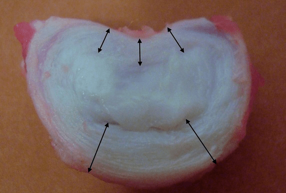
What is the structure of the intervertebral disc?
Each intervertebral disc consists of two different parts:
- outer fibrous ring, consisting of dense fibers that cover the disc from the outside around the circumference;
- The elastic component inside is the nucleus pulposus.
The fibers of the annulus fibrosus are very dense and elastic.Over the years, elasticity is lost and by the age of 60, the fibrous ring becomes stiff.Between the surface of each vertebra above and below and the intervertebral disc itself there is the so-called endplate, that is, the border zone between the vertebra and the intervertebral disc.Thanks to these end plates, the vertebrae grow in height and through them the nucleus pulposus and tissues of the intervertebral disc are nourished by diffusion, since the cartilage of the intervertebral disc is not supplied with blood or innervated.
Healthy intervertebral discs in young people have high metabolism.If you put contrast on a normal plate, then after 20 minutes it will disappear from the plate.
Studies have shown that in adults, the height of each intervertebral disc is approximately:
- 25% of the height of adjacent vertebrae in the cervical region;
- 20% in chest;
- 33% in the lumbar region.
That is, in the lumbar region, the thickness of the intervertebral disc is greatest due to the greatest load.Laboratory studies have shown that a healthy intervertebral disc in a young person can withstand a static compressive load of up to 2.5 tons.At age 70, this figure drops to 110 kg!That is, an “old and dry disc” will cope 22 times worse with transferring load to the sides and maintaining increased pressure within the ring.
Why does this happen?Over time, the thread gradually wears away.It can no longer stretch and just protrude outward, beyond the disc or break.The core stops transmitting and converts the longitudinal load into a radial load.With age, stress gradually builds up inside the discs and their structure changes.If all these processes, carried out in a separate disc, were transferred to the entire spine, then we would have a condition called osteoarthritis in the clinic.Now we can begin to identify.
What is osteonecrosis?
The name of the disease is scary when unclear.The medical suffix “-oz” means proliferation or expansion of certain tissues: hyalinosis, fibrosis.An example is cirrhosis, when connective tissue grows and the functional tissue, hepatocytes, decreases in volume.There may be pathological protein or amyloid deposits that normally should not be present.This storage disease will then be called amyloidosis.There may be significant enlargement of the liver due to fatty degeneration, known as fatty liver disease.
Well, it turns out that with osteoarthritis of the vertebrae, the cartilaginous tissue of the intervertebral discs increases in volume, because “chondros, χόνδρο” translated from Greek into Russian means “cartilage”?No, cartilage disease, or more precisely osteoarthritis, is not a storage disease.There is no actual growth of cartilage tissue in this case;we are just talking about changing the configuration of the intervertebral discs under the influence of many years of physical activity, and we have considered above what happens to each individual disc.The term “osteonecrosis” was introduced into the clinical literature in 1933 by A. Hilderbrandt.
Chondrolysis refers to dystrophic degenerative processes and is part of the normal aging process of intervertebral discs.None of us is surprised that the face of a 20-year-old girl will be slightly different from her face at 70, but for some reason everyone believes that the spine, its intervertebral discs, do not undergo such pronounced temporary changes.Dystrophy is a nutritional disorder, degeneration is a violation of the structure of the intervertebral discs after a long period of dystrophy.
Causes of osteoarthritis and its complications
The main cause of uncomplicated physiological osteoarthritis can be considered the way a person moves: walking upright.Humans are the only species on earth that walk on two legs among all mammals and this is the only way of locomotion.Osteoporosis has become the scourge of humanity, but we have freed our hands and created civilization.Thanks to walking upright (and osteochondrosis), we not only created the wheel, the alphabet, and mastered fire, but you can also sit at home in the warmth and read this article on your computer screen.
Man's closest relatives, the higher primates - chimpanzees and gorillas, sometimes stand up on two legs, but this method of locomotion is only auxiliary for them and most of them still move on all fours.In order for osteoarthritis to disappear, like severe aging of the intervertebral discs, a person needs to change the way they move and remove constant vertical loads from the spine.Dolphins, killer whales, and whales do not get osteochondrosis, and dogs, cows, and tigers do not get it.Their spine is not subjected to long-term static and vertical shock loads because it is in a horizontal state.If humanity goes to sea and the natural means of transportation is scuba diving, osteoarthritis will be defeated.
An upright posture forced the human musculoskeletal system to develop in a direction that protects the skull and brain from shock loads.But intervertebral discs - elastic cushions between vertebrae - aren't the only protection.A person has elastic foot arches, knee cartilage, physiological curves of the spine: two scoliosis and two kyphosis.All this allows you not to "shake" your brain even while running.
Risk factors
But doctors are concerned about risk factors that can be modified and complications of osteonecrosis avoided, which cause pain, discomfort, limited mobility and reduced quality of life.Let's take a look at the risk factors that are often overlooked by doctors, especially in private medical centers.After all, it is much more profitable to constantly treat a person than to identify the cause of the problem, solve it and lose the patient.Here they are:
- the presence of vertical and horizontal flat feet.Flat feet cause the arch of the foot to stop being elastic, and shock forces are transmitted to the spine without softening.The disc is subjected to significant pressure and rapidly collapses;
- overweight and obesity - no comment needed;
- Improper lifting and carrying heavy objects creates uneven pressure on the intervertebral discs.For example, if you lift and carry a bag of potatoes on one shoulder, the heavy load will fall on one edge of the plate and may be excessive;
- physical inactivity and a sedentary lifestyle.It was said above that it is during sitting that the maximum pressure on the intervertebral discs occurs, since a person never sits straight, but always bends "slightly";
- chronic injuries, ice skating, high-intensity weightlifting, contact martial arts, wearing heavy hats, banging your head against low ceilings, wearing heavy clothes, carrying heavy bags in your arms.
General symptoms
The symptoms described below persist outside the local area.These are common symptoms and can exist anywhere.These are pain, movement disorders and sensory disorders.There are also specific phytonutrient disorders or symptoms, such as urinary disorders, but are less common.Let's take a closer look at these signs.
Pain: muscles and roots
Pain can be of two types: radicular and muscular.Radial pain is associated with compression, or pressing on the protrusion or herniation of the disc of the corresponding root at this level.Each nerve root consists of two parts: sensitive and motor.
Depending on the exact location of the hernia and which part of the root has been compressed, there may be sensory or motor disturbances.Sometimes both disorders occur at the same time, manifesting to different degrees.Pain also belongs to sensory disorders, because pain is a special, specific feeling.
Nerve root pain: radiculopathy due to compression
Nerve root pain is familiar to many people;it is called “neuralgia”.Swollen nerve roots will react violently to any shock and the pain will be very intense, similar to an electric shock.She shoots in the arm (from the neck) or in the leg (from the lower back).Such a strong and painful impulse is called lumbago: in the lower back it is lumbago, in the neck it is lumbago, a rarer term.Such radial pain requires forced, analgesic or analgesic positions.Radial pain occurs immediately when coughing, sneezing, crying, laughing or straining.Any shock to a swollen nerve root causes increased pain.
Muscle pain: muscle tonic
But a herniated disc or herniated disc may not compress the nerve root, but when it moves, it damages the nearby ligaments, fascia and deep muscles of the back.In this case, the pain will be secondary, aching, permanent, stiff in the back and such pain is called myofascial.The source of this pain will no longer be the nerve tissue but the muscles.Muscles can respond to any stimulus in only one way: contraction.And if the stimulation is prolonged, the muscle contraction will turn into continuous spasms, which will be very painful.
A characteristic symptom of such secondary myofascial pain will be increased stiffness in the neck, lower back or thoracic spine, the appearance of dense, painful muscle masses - "rollers" next to the spine, that is, next to the spine.In such patients, back pain increases after several hours of "office" work, prolonged immobility, when the muscles practically cannot work and are in a state of spasm.
Sensory disorders
If a protrusion or herniation, or a muscle spasm presses on the sensitive part of the nerve root, various sensory disturbances will occur.They may accompany the pain or may occur individually after the pain has passed.There are also completely painless, but rare, forms of sensory disorders.
Many people know the condition of numbness of the fingertips and toes (reduced sensation or complete anesthesia), reduced skin sensitivity in the form of long stripes and roots.Sometimes paresthesias occur or form, a feeling of “goosebumps”.Typically, sensitivity disorders occur in the feet, fingertips, and toes.Sensory disorders are quite unpleasant, but sensory disorders do not make a person disabled, but movement disorders can lead to this.
Peripheral movement disorders
If a motor neuron or axons that are part of the motor part of the nerve are affected, the muscle will become weak or completely immobile.In the second case, we are talking about complete paralysis, and in the first case - about paralysis.Paralysis is partial paralysis when the muscles do not function at full capacity.
Most often, such disorders appear in the legs, with protrusion and herniation of the lumbar spine.There are motor structures that control the muscles of the legs and feet.Therefore, with complex, progressive lumbar osteoarthritis, the feet can be impacted.It turns inward, people are forced to raise their legs high to walk with the beaten leg, this is called walking, "chicken pose".
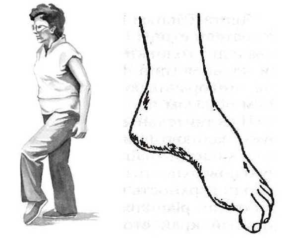
But the whole danger of movement disorders is that they can be isolated and not accompanied by pain.And if a person is “not in pain,” he or she may not see a doctor in time.Therefore, it is important for patients with progressive protrusions and hernias, such as the lumbar region, to periodically walk on their toes and heels and monitor muscle activity.
Local symptoms: main signs
Let's now consider the specific symptoms and syndromes that characterize cervical, thoracic and lumbar osteoarthritis.Let's go from top to bottom, from the neck area down, through the chest area, to the lumbosacral area.
Diagnosis of osteoarthritis
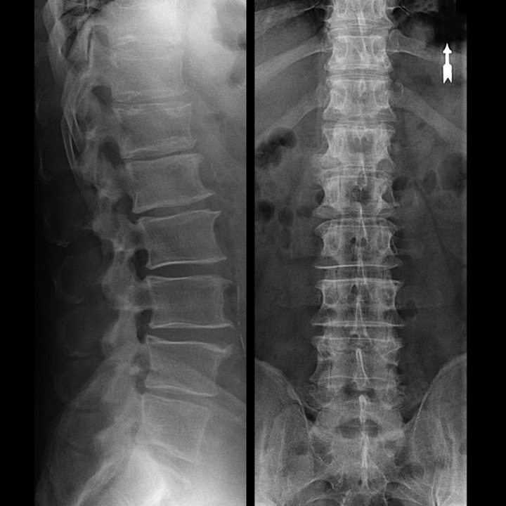
In typical cases, osteoarthritis of the cervical and cervicothoracic spine occurs as described above.Therefore, the main stage of diagnosis remains the identification of the patient's complaints, determining the presence of concomitant muscle spasms by simple palpation of the muscles along the spine.Can the diagnosis of osteoarthritis be confirmed with an X-ray?
An “X-ray” of the cervical spine and even with functional tests of flexion and extension do not show cartilage because their tissue transmits X-rays. Despite this, based on the position of the vertebrae, one can draw general conclusions about the height of the intervertebral discs, the general straightness of the physiological curvature of the neck - scoliosis, as well as the presence of marginal growths on the vertebrae with prolonged irritation of their surfaces by fragile and dehydrated intervertebral discs.Functional tests can confirm the diagnosis of cervical spine instability.
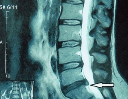
Since the disc itself can only be seen using CT or MRI, magnetic resonance imaging and X-ray computed tomography are indicated to clarify the internal structure of the cartilage and components such as protrusions and herniations.Therefore, with the help of these methods, the diagnosis will be made accurately and the results of the CT scan will be an indication, even a local guide for surgical treatment of hernia in the neurosurgery department.
Treatment of complications of osteoarthritis
Let us repeat once again that osteoarthritis, such as planned aging and dehydration of the intervertebral disc, cannot be cured.You can simply not let things get complicated:
- If you have symptoms of narrowing the height of the disc, you need to exercise properly, not gain weight and avoid bulging and muscle pain;
- if you already have a protrusion, you need to be careful not to let it break the annulus, that is, not to turn the protrusion into a hernia and avoid the appearance of protrusions at multiple levels;
- If you have a hernia, then you need to monitor it dynamically, take regular MRI scans, avoid increasing its size or proceed with treatment with modern minimally invasive surgery, since without exception, all conservative treatments for exacerbation of osteoarthritis leave the hernia and eliminate only temporary symptoms: inflammation, pain, shooting and muscle spasms.
But with the slightest violation of the regimen, heavy lifting, hypothermia, injury, weight gain (in case of back pain), the symptoms return.We will describe how you can cope with the discomfort, pain and limited mobility in your back caused by exacerbation of osteonecrosis and existing protrusion or herniation, secondary to hypertonicity syndrome.
What to do during an exacerbation?
Because there is already acute pain (e.g. in the lower back), you need to follow these instructions at the pre-examination stage:
- completely eliminate physical activity;
- sleep on a hard mattress (orthopedic mattress or hard sofa), eliminating back sagging;
- semi-rigid corsets should be worn to avoid sudden movements and “deformations”;
- You should place a massage pillow with a plastic needle tip on your lower back or use a Lyapko applicator.You need to keep it for 30 - 40 minutes, 2 -3 times a day;
- After that, ointments containing NSAIDs, bee or snake venom ointments can be rubbed into the lower back;
- After rubbing, on the second day you can wrap your lower back with dry heat, such as a belt made from dog hair.
A common mistake is to warm up on the first day.This can be a heating pad, bath procedures.At the same time, the swelling only gets worse and with it pain.You can only warm up after the “most painful point” has passed.Heat then enhances the ability to “reabsorb” the swelling.This usually happens in 2-3 days.
The basis of any treatment is etiotropic therapy (elimination of the cause) and pathological treatment (affecting the mechanism of the disease).It comes with symptomatic treatment.For vertebral pain (caused by problems in the spine), it goes like this:
- To reduce swelling of the muscles and spine, a salt-free diet and restriction of fluid intake are prescribed.You can even give a mild potassium-sparing diuretic pill;
- In the acute phase of lumbar osteoarthritis, short-term treatment can be carried out with intramuscular injections of NSAIDs and muscle relaxants: daily.This will help reduce swelling of nerve tissue, eliminate inflammation and normalize muscle tone;
- in the subacute period, after overcoming maximum pain, no more "injections" should be given, but attention should be paid to restorative drugs, for example, modern drugs of group "B".They effectively restore impaired sensitivity, relieve numbness and paresthesia.
Physical therapy measures continue, it is time to apply exercise therapy for osteoarthritis.Its job is to normalize blood circulation and muscle tone when swelling and inflammation have subsided but muscle spasms have not completely resolved.
Occupational therapy (movement treatment) includes performing therapeutic exercises and swimming.Exercises to treat cervical spondylosis do not target the discs at all but target the surrounding muscles.Its task is to relieve tonic spasms, improve blood flow and normalize venous outflow.This is what leads to decreased muscle tone, reduced severity of pain and stiffness in the back.
Exercises to treat osteoarthritis must be performed after a light warm-up, on warmed muscles.The main therapeutic element is movement, not the degree of muscle contraction.Therefore, to avoid recurrence, the use of weights is not allowed;a gym mat and a gym stick are used.With their help, you can effectively restore range of motion.
Continue rubbing the ointment and use the Kuznetsov applicator.Swimming, underwater massage, and Charcot bathing are shown.It is during the period of remission that magnetic therapy drugs and home physiotherapy are prescribed.
Usually, treatment takes no more than a week, but in some cases, osteonecrosis can present with symptoms so dangerous that surgery and emergency treatment may be necessary.
About Shants collar
In the early stages, in the acute phase, it is necessary to protect the neck from unnecessary movements.Shants collars are great for this.Many people make two mistakes when buying this necklace.They do not choose it according to their size, which is why it does not perform its function and causes discomfort.
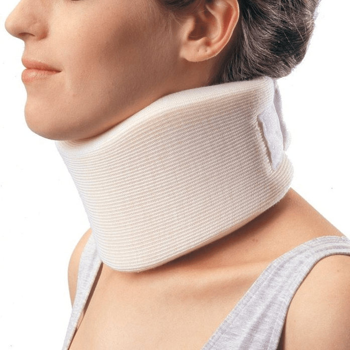
The second most common mistake is wearing it for long-term disease prevention purposes.This leads to weak neck muscles and only causes more problems.As for the collar, there are only two signs that it can be worn:
- Acute pain in the neck, stiffness and pain radiating to the head;
- If you plan to engage in physical work while you are still in good health, this risks “stressing” your neck and making it worse.For example, this is fixing a car when you lie under it or cleaning windows when you need to stretch your arms and take awkward positions.
The collar should not be worn for more than 2–3 days, as wearing it longer can cause venous congestion in the neck muscles, at a time when the patient must activate it.An analogue of the Shants collar for the lower back is a semi-rigid corset purchased at a orthopedic store.
Surgical treatment or conservative measures?
Every patient, after the progression of symptoms, when complications arise, should have an MRI scan and consult a neurosurgeon.Modern minimally invasive operations help to safely remove quite large hernias, do not require a long hospital stay, do not have to stay for several days, do not affect the quality of life, since they are performed using video endoscopy, radiofrequency, modern laser technology or using cold plasma.You can vaporize part of the nucleus and reduce the pressure, reducing the risk of a hernia.And you can completely eliminate the defect, that is, completely eliminate it.
There is no need to fear hernia surgery;This is no longer the previous open surgery of the 80s and 90s of the last century with muscle dissection, blood loss and long recovery time afterward.They are like a small puncture under X-ray control, which is then used using modern technology.
Prevention of osteoarthritis and its complications
Osteoarthritis, including the complex diseases, symptoms and treatments we discussed above, is for the most part not a disease but simply a manifestation of the inevitable aging process and premature “shrinkage” of intervertebral discs.Osteoarthritis needs little to never bother us:
- avoid hypothermia, especially in autumn and spring and fall in winter;
- Do not lift weights and only carry heavy objects with a straight back, in a backpack;
- drink more clean water;
- Don't get fat, your weight should correspond to your height;
- treat flat feet, if present;
- exercise regularly;
- Participate in exercises that help reduce the load on the back (swimming);
- give up bad habits;
- Alternate mental stress with physical activity.After every hour and a half of mental work, the type of activity should be changed to physical;
- You can often have a double-projection X-ray of the lumbar spine or an MRI to see if the herniation is progressing;
By following these simple recommendations, you can keep your back healthy and flexible for life.